

Theoretical Approach
Scientific Paper
This analysis was based on the scientific paper “Modeling the Effect of Multiple Myeloma on Kidney Function” by Julia C. Walk, Bruce P. Ayati and Sarah A. Holstein [1] and his available in https://arxiv.org/pdf/1602.03214.pdf.
Glossary
Multiple Myeloma: Cancer formed in plasma cells, a type of white blood cells [2] [3].
Monoclonal Free Light Chains: Earliest biomarkers in malignant plasma cell-proliferative disorders [4] [5].
Tubulointerstitial fibrosis: Process initiated by the interaction of free light chains (FLCs) and tubule cells, causing inflammation on the kidneys [1]. Regular end-stage for renal diseases [6].
Fibroblasts: Active cells of connective tissue, responsible for synthetize collagen and extracellular matrix [7] [8].
Proteinuria: High concentrations of protein in the urine [9]. Overflow proteinuria can be related with an enlarged production of light chains in multiple myeloma [10].
Content Review
Multiple myeloma is a form of cancer that affects the plasma cells, a type of white blood cells located in bone marrow. When cells become malignant, they overproduce a protein that grows and takes over the bone marrow, blocking the action of healthy cells [11].
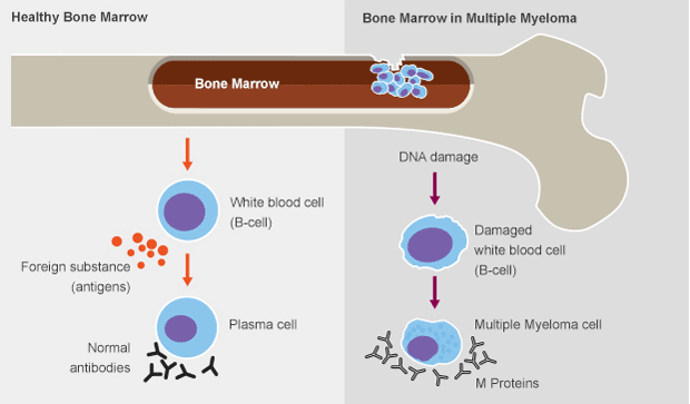
Figure 1:Healthy bone marrow and bone marrow in multiple myeloma. Taken from [12].
Although it has been given more attention to how multiple myeloma affects the bone, it is also important to examine how it affects the kidneys [1]. Fast evolution of tumor cells inhibits the bone rebuilding and they begin to degenerate, which can cause proteinuria, once it is released high amounts of calcium and proteins to the bloodstream, instigating kidneys to overwork and consequent failure [11].
The protein that leads to this damage is called monoclonal immunoglobulin (Ig) light chains (also named M proteins). It is a subunit of antibodies, which are produced to combat infections. Tumor plasma cells overproduce this protein, leading to the most common cause of severe renal failure, tubulointerstitial pathology, resulting from the high concentration of Ig light chains. However, not all the FLCs are toxic to kidneys, appearing this to be related to its structure or protein folder [1].
FLCs can be noxious to proximal tube cells (PTCs) by activating tubulointerstitial fibrosis, an inflammation that causes the excess accumulation of extracellular matrix, which may be replaced for scar tissue – which is largely irreversible. This process leads to the damage of proximal tubule cells and end stage renal disease [1].
Computational Simulation
Algorithm Analysis
The following analysis is based on what is referred and reported in [1]. The following table, transcribed from [1], shows the list of parameters used in the simulation, as well as its meanings and values.
Table 1: List of parameters used in the simulation. Transcribed from [1].
| Parameter | Definition | Value |
|---|---|---|
| γL | FLC growth constant | 0.005 cells-1 days-1 |
| γT | Tumor growth constant | 0.05 days-1 |
| γF | Fibroblast growth constant | 0.004 dL/(mg days) |
| βP | PTC proliferation constant | 0.004 days-1 |
| LS | FLC saturation constant | 400 mg/dL |
| LT | Maximum tumor size | 100 (maximum percentage) |
| µP | PTC natural apoptosis rate | 0.0035 days-1 |
| µL | Natural FLC apoptosis rate | 0.0005 days-1 |
| µF | Natural fibroblast apoptosis rate | 0.0004 days-1 |
| g1 | Strength of inhibited FLC on PTC growth | 1 (dimensionless) |
| g2 | Strength of PTC on its own growth | 1 (dimensionless) |
| g3 | Strength of FLCs on PTC growth | 0.3 (dimensionless) |
| g4 | Strength of PTC on its own death | 0.2 (dimensionless) |
| g5 | Strength of tumor on FLC growth | 0.5 (dimensionless) |
| g6 | Strength of FLC on its own growth | 0.5 (dimensionless) |
The authors choose to use Gompertz model to model the evolution of multiple myeloma [1].
dP/dt = βP (1 - L/LS)+g1 Pg2 - µPP - γFLg3 Pg4 (1)
dL/dt = γL Tg5 Lg6 - µLL (2)
dT/dt = γT Tlog(LT/T) (3)
dF/dt = γF Lg3 Pg4 - µFF (4)
Equation 1, Equation 2, Equation 3 and Equation 4 describe, respectively, the population of proximal tubule cells, the amount of free light chains, the tumor density and the populations of fibroblasts, at time t [1].
The initial conditions presented in the scientific paper for the mathematical model, and used in the simulation, are: P(0) = 400, L(0) = 90, T(0) = 1 and F(0) = 10 [1].
Results Interpretation
In this simulation, the forms of our graphics follow the ones showed in the paper [1]. However, there is a difference in the evolution through time of the graphics, since, in our simulation, it is needed a little greater time interval to reach the same values as in [1].
The behavior of the graphics is consistent to what was expected. As the density of the tumor cells increases, the FLCs amount and fibroblasts population rises and the population of proximal tubule cells falls [1].
References
[INTRO IMAGE] Boonyarit Cheunsuchon, “Myeloma kidney,” Renal Pathology Review, 8 May 2013. [Online]. Available: http://renalpathologyreview.blogspot.pt/2013/05/myeloma-kidney.html. [Acedido em 25 August 2016].
[1] Julia C. Walk, Bruce P. Ayati, Sarah A. Holstein, “Modeling the Effects of Multiple Myeloma on Kidney Function,” arXiv:1602.03214v1 [q-bio.TO], 2016.
[2] Mayo Clinic Staff, “Multiple myeloma,” Mayo Foundation for Medical Education and Research, [Online]. Available: http://www.mayoclinic.org/diseases-conditions/multiple-myeloma/basics/definition/con-20026607. [Acedido em 29 August 2016].
[3] “What is multiple myeloma?,” American Cancer Society, [Online]. Available: http://www.cancer.org/cancer/multiplemyeloma/detailedguide/multiple-myeloma-what-is-multiple-myeloma. [Acedido em 29 August 2016].
[4] Ellen Jenner, “Serum free light chains in clinical laboratory diagnostics.,” Clinica Chimica Acta, vol. 427, pp. 15-20, 2014.
[5] Zsuzsa Végh, Szabolcs Ottó, Sándor Eckhardt, “Monoclonal free light chains in urine and their significance in clinical diagnostics: are they really tumor markers?,” Journal of clinical laboratory analysis, vol. 4, nº 6, pp. 443-448, 1990.
[6] Masayuki Iwano, Eric G. Neilson, “Mechanisms of tubulointerstitial fibrosis.,” Current opinion in nephrology and hypertension, vol. 13, nº 3, pp. 279-284, 2004.
[7] Dr Ananya Mandal, MD, “What are Fibroblasts?,” News-Medical.ne, [Online]. Available: http://www.news-medical.net/health/What-are-Fibroblasts.aspx. [Acedido em 29 August 2016].
[8] The Editors of Encyclopædia Britannica, “Fibroblast,” Encyclopædia Britannica, Inc., [Online]. Available: https://www.britannica.com/science/fibroblast. [Acedido em 28 August 2016].
[9] Anuja P. Shah, MD, “Proteinuria,” Merck Sharp & Dohme Corp., [Online]. Available: https://www.merckmanuals.com/professional/genitourinary-disorders/symptoms-of-genitourinary-disorders/proteinuria. [Acedido em 29 August 2016].
[10] Edgar V Lerma, “Proteinuria,” WebMD LLC, [Online]. Available: http://emedicine.medscape.com/article/238158-overview. [Acedido em 29 August 2016].
[11] Written by Vanessa Bates Ramirez. Reviewed by George T. Krucik., “The Link Between Multiple Myeloma and Kidney Failure,” 17 November 2014. [Online]. Available: http://www.healthline.com/health/cancer/multiple-myeloma-kidney-failure#Overview1. [Acedido em 30 August 2016].
[12] “What is Multiple Myeloma?,” Multiple Myeloma Research Foundation, [Online]. Available: https://www.themmrf.org/multiple-myeloma/what-is-multiple-myeloma/. [Acedido em 30 August 2016].
[13] Armando Hasudungan, “Medicine - Multiple Myeloma,” ÝouTube, 26 July 2015. [Online]. Available: https://www.youtube.com/watch?v=ghvoKhpAc64. [Acedido em 27 August 2016].